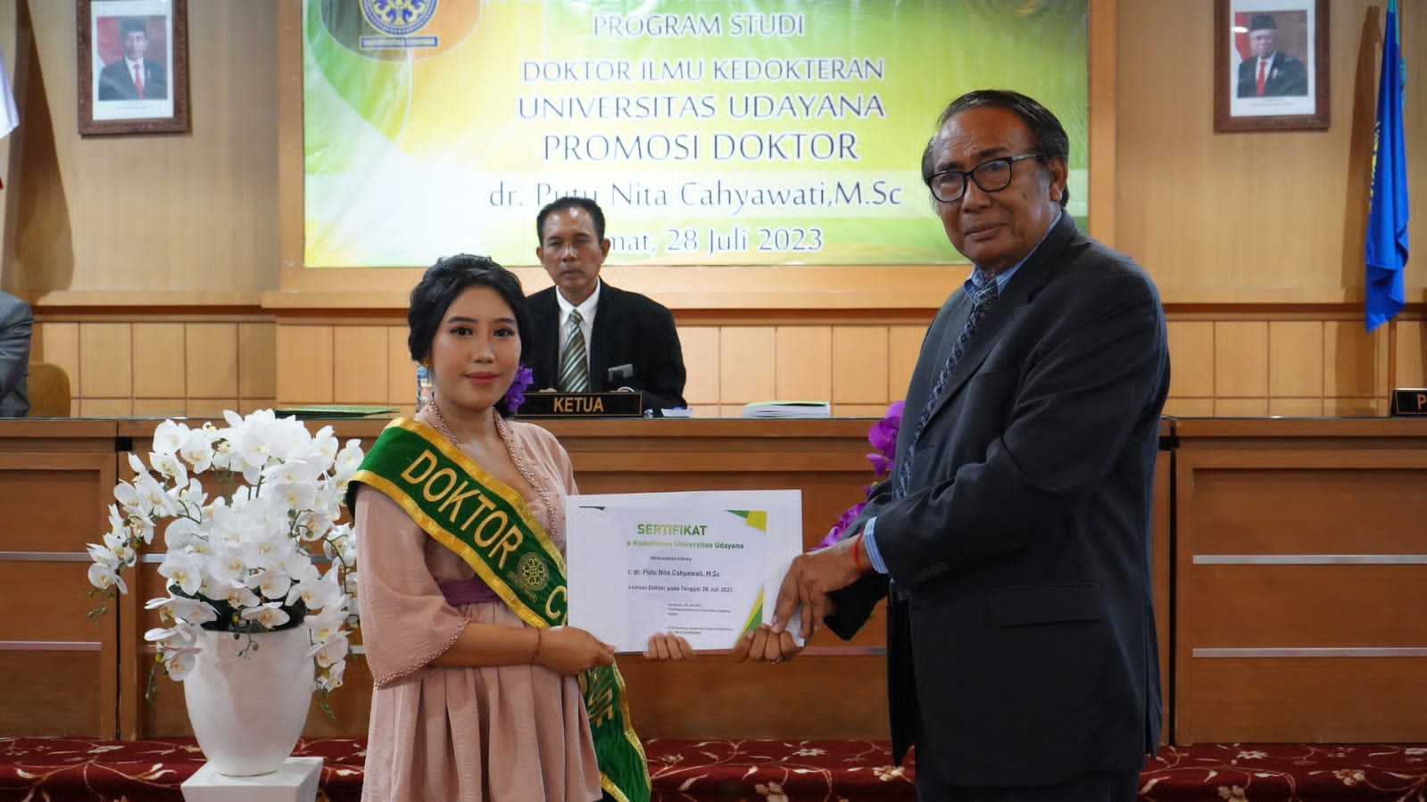
Producing Theories Related to Simvastatin Mechanisms in Reducing Kidney Fibrosis, Nita Cahyawati Gets Doctor of Medicine
Taking place in the Postgraduate Meeting Room, Denpasar Postgraduate Building, a Doctoral Promotion exam was held with the promovenda candidate, dr. Putu Nita Cahyawati, M.Sc, with the dissertation title "Effect of Simvastatin on Tumor Necrosis Factor-Alpha Expression, Alpha-Smooth Muscle Actin Expression, and Degree of Kidney Tissue Interstitial Fibrosis in Mice Model of Chronic Kidney Failure" (26/7/2023)
Chronic kidney failure (CKD) is a progressive disease whose prevalence continues to increase every year. Studies in recent years have reported that statins are able to prevent the progression of kidney damage, but the repair mechanism is still unclear, fragmented, and controversial. This study aims to clarify the theory regarding the mechanism of simvastatin in the prevention of renal fibrosis in the subtotal nephrectomy model, through analysis of TNF-α expression, myofibroblast cell expansion, and collagen deposition in the renal interstitium.
This research is a true experimental study (randomized post-test only control group design). The research sample consisted of 20 white mice (Mus musculus L.), Swiss line, aged 3-4 months, weighing 30-40 grams. Samples were randomly grouped into four treatment groups: control (K, n = 5), simvastatin dose 5.2 mg/kgBB (P1), simvastatin dose 10.4 mg/kgBB (P2), and simvastatin dose 20.8 mg/kg kgBB (P3). TNF-α expression was assessed using real time-PCR, α-SMA expression was assessed by immunohistochemical examination, and interstitial fibrosis was assessed using Picrosirus Red. Data analysis used Tukey's one-way ANOVA and post hoc tests with a significance value of p< 0.05. Path analysis is used to determine the pattern of relationships between variables. The results showed that the control group (K) was the group with the highest mean serum creatinine level (1.07 ± 0.43 mg/dL) compared to the other treatment groups (p < 0.05).
Administration of simvastatin at doses of 5.2 mg/kg, 10.4 mg/kg, and 20.8 mg/kg resulted in 2 to 4 lower expression of TNF-α in the kidney tissue than controls. There was no significant difference in the expression of TNF-α in the kidney tissue at the three dose levels. Administration of simvastatin at doses of 5.2 mg/kg, 10.4 mg/kg, and 20.8 mg/kg also resulted in lower α-SMA expression and interstitial fibrosis of kidney tissue compared to controls. There was a significant difference between the expression of α-SMA in the kidney tissue in the 5.2 mg/kgBB (P1) simvastatin group and the 20.8 mg/kgBB (P3) simvastatin group, and there was a significant difference between kidney tissue fibrosis in the 5-dose simvastatin group. 2 mg/kgBW (P1) with a dose of 10.4 mg/kgBW (P2) and a dose of 20.8 mg/kgBW (P3). The path analysis results showed that the ability of simvastatin to trigger a lower degree of renal interstitial fibrosis in the CRF animal model was not directly due to lower TNF-α expression, but through lower α-SMA expression.
The conclusion of this study is that simvastatin causes TNF-α expression, α-SMA expression, and a lower degree of fibrosis than the control as the dose is increased.
The novelty of this study is to produce a theory regarding the mechanism of simvastatin in reducing renal fibrosis in white mice (Mus musculus L.) undergoing subtotal nephrectomy procedures. This study found that simvastatin was able to reduce kidney fibrosis and improve kidney function because of its ability to cause lower TNF-α expression, lower α-SMA expression which reflects the expansion of myofibroblast cells, and lower collagen deposition in the renal interstitium. Path analysis showed that simvastatin's ability to cause a lower degree of interstitial fibrosis was not directly caused by the low expression of TNF-α, but due to the low expression of α-SMA.
The exam was led by the Deputy Dean for General Affairs and Finance of FK Unud, Dr. dr. Made Sudarmaja, M.Kes., with a team of examiners:
1. Dr. dr. I Made Bakta, Sp.PD-KHOM (Promoter)
2.Dr. dr. Bagus Komang Satriyasa, M.Repro (Co-promoter I)
3. drh. I Nyoman Mantik Astawa, Ph.D (Co-promoter II)
4. dr. Ketut Tirtayasa, MS., AIF
5. Prof. Dr. dr. I Made Jawi, M. Kes
6. Dr. Ir. Ida Bagus Putra Manuaba, M. Phil
7. Dr. dr. I Putu Gede Adiatmika, M.Kes
8. Dr. dr. I Wayan Putu Sutirta Yasa, M.Si
9. Prof. dr. I Gusti Made Aman, Sp.FK
While academic invitations are:
1.Dr. dr. Ni Made Linawati, M.Sc
2.Dr. dr. Agung Wiwiek Indrayani, M.Kes
3. Desak Ketut Ernawati, S.Si, Apt., P.G.Pharm., M.Pharm., Ph.D.
4.Dr. dr. Putu Ayu Asri Damayanti, M.Kes
5.Dr. dr. Dewa Ayu Agus Sri Laksemi, M.Sc
In this exam, Dr. dr. Putu Nita Cahyawati, M.Sc, was declared the 393rd graduate of the Doctoral Degree, Faculty of Medicine from Udayana University with honors (Cumlaude).


